Nanomaterials for Sample Preparation
用于生物样本制备的纳米材料
收缩诱导多级纳米结构氧化硅

The heat-shrinking process leads to a hierarchical structure of microscale polyolefin folds layered with nanoscale silica lamella that can be fine-tuned via oxide deposition thickness. SEM analysis was performed on Nanobind with: a) 2 nm, b) 20 nm, c) 50 nm, d) 100 nm, e) 150 nm, and f) 200 nm of deposited silicon dioxide.
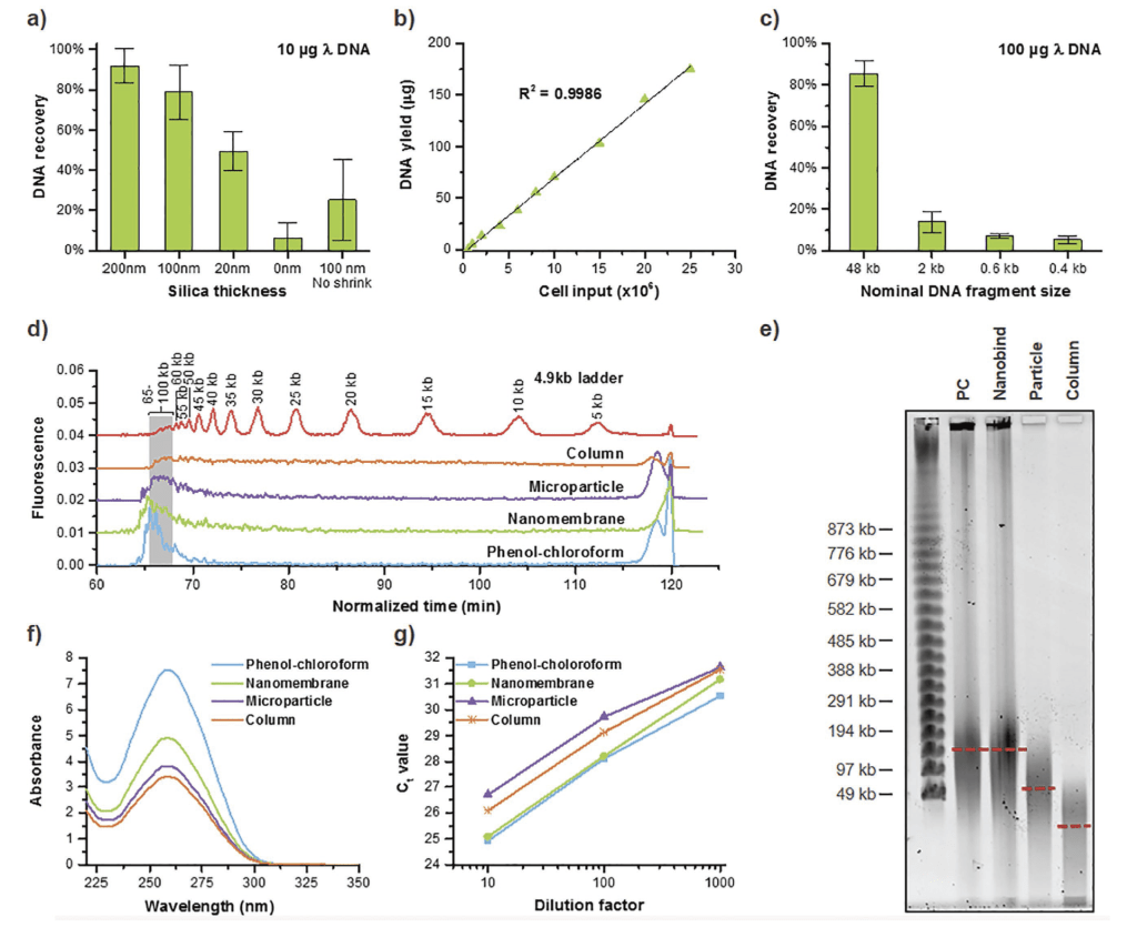
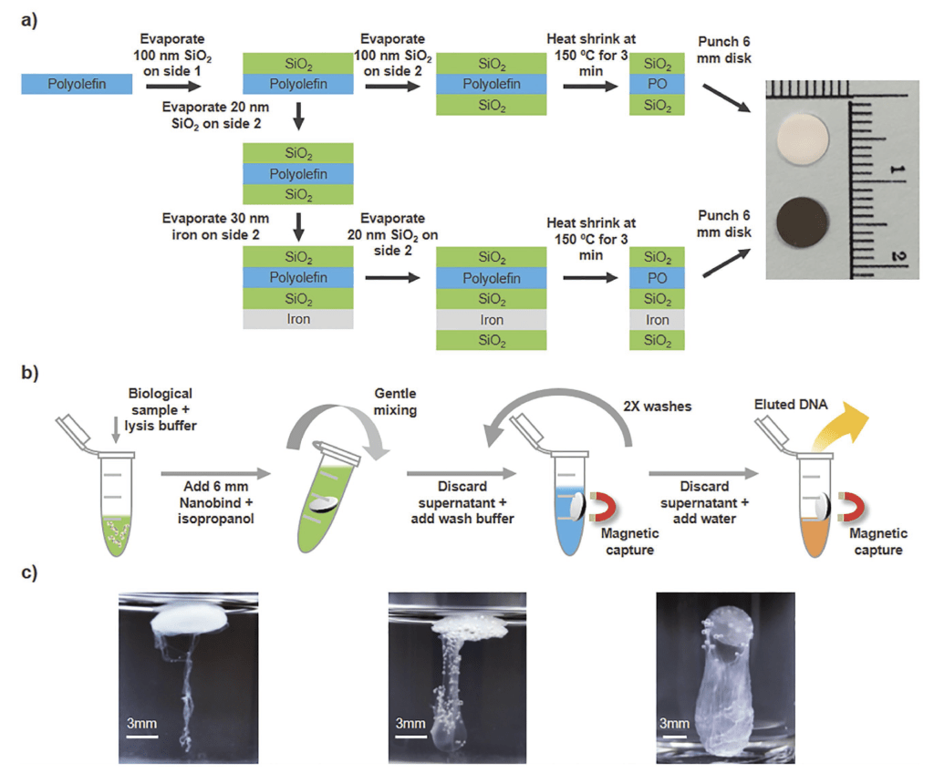
Yi invented NanoBind Technology, a liquid/solid two-phase DNA extraction technology capable of isolating high-purity DNA molecules with ultrahigh molecular weight that are ideally suited for single-molecule long-read sequencing based on shrink-induced hierarchical silica nanofilm. This technology was successfully commercialized by Circulomics Inc. which was acquired by Pacific Biosciences. Nanobind has already become the standard sample preparing technology for third-generation DNA sequencing platforms.
张翼开发了一套通过收缩诱导制备具备多级微纳结构的二氧化硅薄膜的即时,适用于大规模制备此类纳米材料。基于微纳多级二氧化硅膜的固液双相大分子量DNA萃取技术已完成技术转换,并被第三代测序领跑企业Pacific Biosciences收购,相关技术产品目前已成为第三代长读测序的样本制备标准方法。
A Simple Thermoplastic Substrate Containing Hierarchical Silica Lamellae for High-Molecular-Weight DNA Extraction, Advanced Materials, 2016, 28, 10630–10636
Nanomaterials for Biosensing
用于生物传感的纳米材料
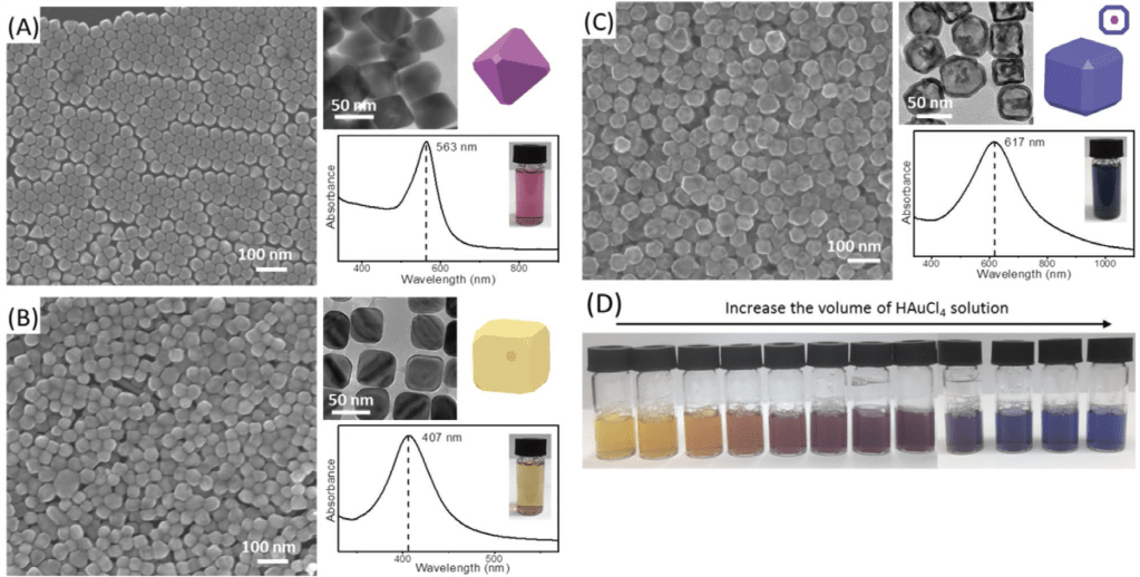
SEM images, TEM images (upper left inset), geometric models (upper right inset), and ultravioletvisible (UV–vis) spectrum (lower inset) of A) Au NPs, B) Au@Ag coreshell NPs, and C) Au/Ag hollow NPs. D) Vials containing Au/Ag NPs prepared by reacting Au@Ag core-shell NP solution with different volumes of a 20 mm HAuCl4 solution.
SEM图像,TEM图像(左上插图),几何模型(右上插图)紫外线-可见光(UV-VIS)光谱(下插图)。(A)金纳米粒子。(B)金@银核-壳纳米粒子。(C)金/银空心纳米粒子。(D)含有金/银纳米粒子的小瓶,由金@银核-壳纳米粒子溶液与不同体积的20 mM的HAuCl4溶液。

Color combinations using NPs of three primary colors. A) Combination of NPs of three primary colors results in NP mixtures of unique secondary colors. B) Color palette by mixing primary NPs at various ratios. Droplets in the first row are mixtures of yellow and magenta NPs, droplets in the second row are mixtures of yellow and cyan NPs, and droplets in the third row are mixtures of magenta and cyan NPs. The ratios of the two NPs in the droplet from left to right are 10:0, 9:1, 8:2, 7:3, 6:4, 5:5, 4:6, 3:7, 2:8, 1:9, and 0:10, respectively. C) FITC-labeled dsDNA target detected by single primary NPs and mixture of NPs.
使用三原色纳米粒子的颜色组合。(A)将三原色的纳米粒子组合,形成独特的次级颜色的纳米粒子混合物(B)以不同比例混合三原色纳米粒子的调色板。第一行液滴是黄色和洋红色纳米粒子的混合物,第二行液滴是黄色和青色纳米粒子的混合物,第三行液滴是洋红色纳米粒子和青色纳米粒子的混合物。液滴中两个纳米粒子的比例从左到右分别是10:0,9:1,8:2,7:3,6:4,5:5,4:6,3:7,2:8,1:9,0:10(C)FITC标记的dsDNA目标物分别被单一纳米粒子与混合的纳米粒子所检测。
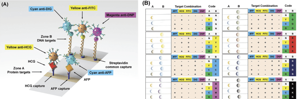
Two-parameter multiplexing based on spatial separation and color co-localization. A) Detection schematics. Cyan, magenta, and yellow NPs are represented by spheres of their respective colors. B) Multiplexed detection of all 32 possible target combinations. Target combinations were reported by the location and the color of the test spot. Co-localization of multiple primary NPs resulted in unique secondary colors.
基于空间分离和颜色共定位的双参数多重检验。(A)检测原理图。青色,洋红色,黄色纳米粒子由其各自颜色的球体表示。(B)对所有32种可能的目标组合进行多重检验。目标组合由通过测试点的位置和颜色来报告。多个主要纳米粒子的共定位产生了独特的次级颜色。
Multi-Color Au/Ag Nanoparticles for Multiplexed Lateral Flow Assay Based on Spatial Separation and Color Co-Localization, Advanced Functional Materials, 2021, 2109553.
